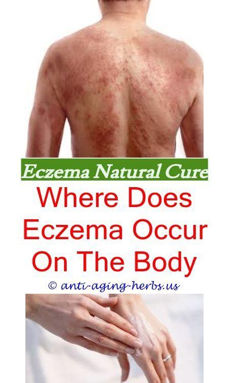
Few sensory experiences are as exhausting as the relentless itch of an eczema flare. It starts as a subtle prickling beneath the epidermis, rapidly escalating into an intense urge to scratch. However, scratching only tears the fragile stratum corneum, allowing environmental irritants, microbes, and allergens to invade deeper tissue and ignite further cutaneous inflammation—a vicious clinical pattern known as the itch-scratch cycle.
Affecting over 31 million children and adults in the United States alone, eczema is far more than superficial “dry skin.” It represents a complex chronic dermatological disorder rooted in epidermal barrier impairment, immune dysregulation, and microbiome imbalance. When your skin barrier is compromised, it loses moisture while inviting opportunistic microbes like Staphylococcus aureus to colonize the surface.
Understanding why your skin barrier fails—and how to biologically reconstruct it from both the outside in and the inside out—is the key to achieving durable, steroid-sparing remission. In this clinical guide, we explore the distinct types of eczema, dissect the cellular biology of barrier dysfunction, review high-yield botanical interventions, and present an actionable relief blueprint.
📋 Medical Summary & Clinical Key Takeaways
- Barrier Pathology: Eczema (atopic dermatitis) stems primarily from epidermal barrier breakdown—frequently involving FLG (filaggrin) gene mutations, diminished ceramide synthesis, and elevated transepidermal water loss (TEWL).
- Immune Activation: A disrupted lipid barrier permits allergen and microbial penetration, triggering Th2-polarized cytokine cascades (IL-4, IL-13) and secondary Staphylococcus aureus colonization.
- First-Line Protocol: Rebuilding structural integrity requires the clinical “Soak and Seal” hydration method, physiological 3:1:1 lipid replenishment, and strict avoidance of harsh surfactant detergents.
- Clinical Red Flags: Honey-colored crusting, rapidly spreading punched-out vesicles (eczema herpeticum), or systemic fever demand immediate physician-directed medical evaluation.

What Exactly Is Eczema? The 7 Primary Types
The medical term eczema originates from the Greek word ekzein, meaning “to boil over”—a vivid portrayal of inflamed, vesicular skin. In clinical dermatology, “eczema” is an umbrella term encompassing several distinct inflammatory conditions. Establishing an accurate diagnosis is crucial, as each presentation responds to different environmental modifications:
1. Atopic Dermatitis (AD)
The most common and chronic form, atopic dermatitis typically begins in infancy or early childhood and often coexists with asthma and allergic rhinitis (the “atopic march”). It primarily targets flexural areas such as the creases of elbows and behind the knees, presenting with severe xerosis, intense pruritus, and lichenified plaques. For chronic itching without active weeping, see our comprehensive protocol on dry, itchy skin relief and barrier restoration.
2. Contact Dermatitis
Triggered when external substances damage the skin barrier. It divides into irritant contact dermatitis (caused by harsh detergents, industrial solvents, or repeated handwashing) and allergic contact dermatitis (a delayed Type IV cell-mediated hypersensitivity reaction to allergens such as nickel, fragrance mixes, or topical preservatives).
3. Dyshidrotic Eczema (Pompholyx)
Characterized by intensely itchy, deep-seated, tapioca-like vesicles along the edges of fingers, palms, and soles. Flares are frequently exacerbated by excessive perspiration, emotional stress, wet work, or contact sensitivities to metals.
4. Nummular Eczema (Discoid Eczema)
Presents as well-defined, coin-shaped (nummular) circular lesions that often crust, weep, and itch intensely. It is commonly triggered by severe winter xerosis, insect bites, or localized skin trauma, and is easily misidentified as fungal ringworm.
5. Seborrheic Dermatitis
Localizes to sebum-rich regions including the scalp (“cradle cap” in infants), eyebrows, nasolabial folds, and upper chest. Driven by inflammatory reactions to Malassezia yeast, it manifests as greasy, yellowish flakes over an erythematous base.
6. Stasis Dermatitis
Also termed gravitational dermatitis, this form occurs on the lower legs of individuals with chronic venous insufficiency. Incompetent venous valves cause fluid pooling and microvascular hypertension, resulting in skin scaling, hyperpigmentation, and aching.
7. Neurodermatitis (Lichen Simplex Chronicus)
A self-perpetuating localized disorder where chronic rubbing or scratching transforms a small irritation into a thick, leathery, hyperkeratotic plaque with exaggerated skin markings, often provoked by heightened nervous tension.
The Biology of a Flare: Why Does the Skin Barrier Fail?
To successfully treat eczema, you must understand the architecture of the stratum corneum—the outermost shield of your skin. Dermatologists describe this structure using the classical “brick and mortar” model: corneocytes (flattened dead skin cells) serve as the bricks, surrounded by a dense matrix of intercellular lipids (ceramides, cholesterol, and free fatty acids) acting as mortar.
In atopic dermatitis, this architectural shield fails due to a convergence of genetic and biochemical defects:
- Filaggrin (FLG) Deficiency: Filaggrin is a vital structural protein that binds keratin fibers within corneocytes and degrades into Natural Moisturizing Factor (NMF). Landmark dermatological trials published in The Journal of Allergy and Clinical Immunology (PubMed PMID: 18454223) demonstrate that genetic loss-of-function FLG mutations significantly destabilize the stratum corneum, precipitating marked transepidermal water loss (TEWL) and widespread hyper-reactivity.
- Lipid Disruption & Ceramide Depletion: Eczematous skin suffers from a marked deficiency in ceramides (particularly Ceramide 1 and Ceramide 3). Without adequate lipids, microscopic gaps emerge between cells, allowing moisture to evaporate and foreign environmental irritants to penetrate deep into living tissue.
- Th2 Immune Skewing: When antigens cross the fractured stratum corneum, epidermal keratinocytes release alarmins (TSLP, IL-25, IL-33). These signal naive immune cells to differentiate into T-helper 2 (Th2) cells, pumping out inflammatory cytokines (IL-4, IL-13, IL-31). Interleukin-31 directly stimulates cutaneous sensory nerve fibers, inducing intense pruritus.
- Microbiome Dysbiosis & Staph Colonization: Healthy skin harbors a rich, protective microbial ecosystem. In active eczema lesions, Staphylococcus aureus expands dramatically—colonizing over 90% of atopic skin. S. aureus secretes delta-toxins and proteases that further cleave barrier junctions and amplify local inflammation.

Common Hidden Triggers That Provoke Flares
While genetic susceptibility provides the dry kindling, environmental and internal triggers strike the match. Systematically identifying and removing these triggers is essential for long-term comfort:
- Harsh Surfactants & Alkaline Soaps: Conventional bar soaps feature a high pH (9.0–10.0), which destroys the skin’s naturally acidic mantle (pH 4.5–5.5), neutralizing antimicrobial lipids and activating inflammatory kallikrein proteases.
- Synthetic Fragrances & Preservatives: Artificial fragrances, parabens, and release agents like methylisothiazolinone (MI) in laundry detergents and lotions represent major contact sensitizers.
- Airborne Allergens & Climate Extremes: Dust mite feces, pet dander, pollen, dry winter heating, and sweltering summer sweat rapidly aggravate pruritus. Because airborne allergens trigger systemic histamine release, explore our guide on the underlying immune causes of allergies.
- Dietary Antigens & Gut Permeability: In pediatric patients and certain adults, common foods like dairy, gluten, eggs, and soy can exacerbate cutaneous flares. Systemic inflammation is deeply intertwined with gut microbiome integrity; review our clinical guide on gut health and mucosal barrier repair.
- Psychological Stress: Emotional tension activates the hypothalamic-pituitary-adrenal (HPA) axis, stimulating systemic cortisol and substance P release, which destabilizes cutaneous mast cells.
The 5-Step Evidence-Based Natural Relief Blueprint
Rebuilding your skin barrier requires shifting away from temporary suppression and focusing on biological restoration. This 5-step clinical protocol targets external barrier repair and internal balance:
Step 1: Master the Clinical “Soak and Seal” Method
Bathing is therapeutic when done correctly. Soak in lukewarm (never steaming hot) water for 10–15 minutes. Avoid scrubbing with washcloths or loofahs. Step out and gently pat the skin with a soft cotton towel, leaving the skin slightly damp. Within 3 minutes (“the 3-minute rule”), apply a rich, fragrance-free occlusive ointment or ceramide cream over every square inch to trap moisture before evaporation occurs.
Step 2: Restore the 3:1:1 Physiologic Lipid Ratio
Thin, watery lotions contain high alcohol and water content, which paradoxically dehydrates damaged skin through evaporation. Choose rich lipid formulations containing a physiologic ratio of ceramides, cholesterol, and free fatty acids (ideal ratio 3:1:1). Pure virgin coconut oil offers additional clinical benefit: clinical trials confirm that topical virgin coconut oil suppresses Staphylococcus aureus colonization while delivering deep emollience.
Step 3: Harness Targeted Botanical Anti-Inflammatories
Clinical botany offers potent, scientifically validated topical extracts that soothe pruritus without steroid thinning:
- Colloidal Oatmeal (Avena sativa): Approved by the FDA as an over-the-counter skin protectant, colloidal oatmeal contains unique polyphenols called avenanthramides. Research demonstrates that avenanthramides dramatically downregulate NF-kB activation, suppressing histamine release and relieving itch (PubMed PMID: 25607907).
- Organic Cold-Pressed Neem Oil: Revered in Ayurvedic medicine, neem seed oil delivers azadirachtin and nimbin, exhibiting broad-spectrum antimicrobial and anti-inflammatory properties against secondary skin pathogens.
- Diluted Apple Cider Vinegar Soaks: Adding 1–2 cups of raw apple cider vinegar to a warm bath helps restore the acidic pH mantle of the skin; learn more about topical uses in our guide on apple cider vinegar for wellness.
Step 4: Heal the Gut-Skin Axis from Within
Up to 70% of human immune tissue resides in the gut-associated lymphoid tissue (GALT). Intestinal permeability enables microbial endotoxins to trigger cutaneous flares. Supplementing with clinically studied probiotic strains (especially Lactobacillus rhamnosus GG), zinc, and omega-3 fatty acids (EPA/DHA) reduces systemic Th2 inflammatory signaling.
Step 5: Create a Hypoallergenic Home Sanctuary
Eliminate environmental irritants. Switch to fragrance-free liquid laundry detergents, wear loose garments made from 100% organic cotton, bamboo, or silk, and run a cool-mist ultrasonic humidifier to maintain indoor humidity between 45% and 55% during dry winter months.
Eczema Relief Solutions Comparison Table
Compare the mechanisms, clinical utilities, and safety considerations of leading natural barrier repair options:
| Intervention | Primary Mechanism | Best Clinical Application | Safety & Application Tip |
|---|---|---|---|
| Colloidal Oatmeal | Avenanthramides block NF-kB and histamine signaling | Acute itching, generalized body flares, soothing baths | Ensure USP cosmetic-grade; soak 15 min, pat dry |
| Ceramide Complex (3:1:1) | Replaces missing stratum corneum lipid mortar | Daily baseline barrier repair, preventing water loss | Apply within 3 minutes of bathing while damp |
| Virgin Coconut Oil | Lauric acid disrupts microbial cell membranes | Mild atopic dermatitis, secondary Staph inhibition | Patch test first; avoid on acne-prone facial skin |
| Raw Neem Oil | Nimbin & triterpenoids deliver broad antimicrobial action | Stubborn discoid patches, secondary pathogen risk | Dilute in carrier oil (jojoba/argan) due to strong odor |
| Probiotic Therapy | Modulates GALT and decreases circulating Th2 cytokines | Chronic recurrent flares, pediatric atopic history | Select multi-strain formulations with L. rhamnosus |
Curated Eczema Relief Essentials
If you or your family members are seeking clean, clinically verified, non-irritating topical solutions to protect and heal damaged skin, these three gold-standard formulations provide exceptional barrier defense:
Vanicream Moisturizing Cream
The gold standard for ultra-sensitive skin. Free of dyes, fragrance, masking fragrance, lanolin, parabens, and formaldehyde releasers. Awarded the National Eczema Association Seal of Acceptance.
Aveeno Baby Eczema Therapy
Formulated with natural colloidal oatmeal and essential ceramides. Clinically proven to soothe itch, irritation, and dryness while restoring the protective epidermal lipid barrier.
TheraNeem Organic Neem Oil
Certified organic, cold-pressed virgin neem seed oil. Packed with essential fatty acids and antioxidants to soothe severe irritation, calm redness, and nourish distressed skin tissues.
Red Flags: When to Seek Immediate Medical Evaluation
While most eczema flares can be managed effectively with diligent barrier care, compromised skin is vulnerable to secondary infections that require prompt medical evaluation:
- Eczema Herpeticum: A serious, potentially vision-threatening viral infection caused by Herpes Simplex Virus (HSV) spreading across broken eczematous skin. Presents as clusters of intensely painful, fluid-filled, dome-shaped blisters that crust into punched-out erosions, accompanied by fever and lymphadenopathy. Requires emergency oral or IV acyclovir.
- Bacterial Impetigo: When Staphylococcus or Streptococcus bacteria colonize lesions, leading to honey-colored crusting, yellow oozing pus, localized heat, red streaks, and swelling. Requires targeted topical or oral antibiotics.
- Systemic Signs: Sudden onset of high fever, chills, dizziness, lethargy, or rapidly spreading redness indicates a potential bloodstream infection (cellulitis or sepsis).
- Treatment Failure: If severe flares fail to improve within 7–10 days of diligent emollient care, consult a board-certified dermatologist for formal patch testing or systemic biologic therapy.
Related Clinical & Dermatological Guides
- Dry, Itchy Skin Relief: Causes, Lipid Barrier Science & Proven Natural Remedies
- Naturally Clear Skin: The Science of Acne, Gut-Skin Axis & Proven Home Remedies
- Athlete’s Foot (Tinea Pedis): Antifungal Science & Proven Home Remedies
- The Dirty 30: Toxic Beauty Ingredients to Avoid for Cleaner, Safer Skin
- The Complexity of the Gut: Clinical Microbiome & Mucosal Barrier Repair
Frequently Asked Questions (FAQ)
Is eczema contagious to other people?
No. Eczema is entirely non-contagious. You cannot catch eczema from touching an affected person, nor can you transmit it through shared pools, bedding, or towels. It is an internal genetic, immune, and barrier disorder.
Can eczema be permanently cured?
While conventional medicine does not recognize a permanent cure, long-term clinical remission is entirely achievable. By identifying individual biochemical triggers, repairing the stratum corneum with physiological lipids, balancing the microbiome, and managing stress, most individuals live flare-free.
What is Topical Steroid Withdrawal (TSW)?
Topical Steroid Withdrawal (or Red Skin Syndrome) is a debilitating condition that can occur after prolonged, continuous application of high-potency topical corticosteroids followed by abrupt cessation. Symptoms include burning erythema, thermal dysregulation, severe flaking, and rebound flares. Always taper steroids under medical supervision and prioritize non-steroidal barrier repair.
Does swimming in chlorinated pools help or harm eczema?
Chlorinated pool water acts as a mild bleach bath, which can significantly decrease Staphylococcus aureus colonization on eczematous skin. However, chlorine also strips natural sebum. To swim safely, apply a thin protective layer of ointment beforehand, rinse thoroughly with fresh water immediately afterward, and re-apply moisturizer within 3 minutes.
Medical Disclaimer: This article is intended strictly for educational and informational purposes and does not constitute individualized medical advice, diagnosis, or treatment. Always consult a qualified healthcare provider or board-certified dermatologist regarding medical conditions or before initiating new therapeutic regimens.