
Coronary artery disease (CAD) remains the single leading cause of mortality across the developed world, responsible for one out of every five cardiovascular deaths. Yet the most insidious aspect of this condition is its profound silence: atherosclerosis does not develop overnight. It is a slow, painless vascular remodeling process that takes decades to accumulate. An individual can live forty, fifty, or sixty years with high energy, passing standard yearly physicals, completely unaware that a necrotic core of cholesterol plaque is steadily narrowing the main arterial pathways supplying their myocardium. Often, the very first symptom someone experiences is not a subtle warning ache, it is a catastrophic myocardial infarction.
Understanding the silent biology of coronary artery disease is the most empowering preventative health step you can take. When you comprehend how arterial micro-tears attract circulating atherogenic particles, how chronic vascular inflammation destabilizes fibrous lesions, and how modern non-invasive imaging can detect early calcification years before symptoms manifest, you gain the ability to intervene decisively. Here is your evidence-based medical guide to understanding coronary artery disease, recognizing silent and atypical symptoms, and applying proven lifestyle habits to halt arterial plaque.
The Decades-Long Timeline: How Coronary Plaque Silently Develops
To visualize coronary artery disease, imagine the arterial wall not as an inert plastic tube, but as a living, highly reactive organ called the vascular endothelium. Your heart beats approximately 100,000 times each day, propelling pressurized blood through three main coronary arteries: the left anterior descending (LAD), the circumflex, and the right coronary artery (RCA). Over a lifetime, this constant hemodynamic pulsatility interacts with systemic metabolic risk factors across four distinct stages:
Stage 1: Endothelial Injury and Apolipoprotein Infiltration
In your twenties and thirties, high blood pressure, circulating toxic chemicals from tobacco, chronic hyperglycemia, and systemic inflammation cause microscopic abrasions in the endothelial monolayer. Circulating apolipoprotein B-containing particles, chiefly low-density lipoproteins (LDL) and Lipoprotein(a), penetrate these microscopic fissures into the sub-endothelial space (the tunica intima). Trapped inside the arterial wall, these lipids undergo oxidative modification, triggering a chemical distress signal.
Stage 2: Foam Cells and Fatty Streaks
In response to oxidized LDL, the vascular endothelium expresses adhesion molecules that recruit circulating monocytes into the vessel wall. These monocytes differentiate into macrophages, which ravenously engulf the oxidized lipids via scavenger receptors. As macrophages become bloated with cholesterol esters, they transform into “foam cells.” Aggregations of foam cells form macroscopic “fatty streaks” along the inner arterial walls. At this stage, the condition is completely painless and invisible on standard resting electrocardiograms (ECGs).
Stage 3: The Fibrous Cap and Calcification
Over your forties and fifties, the body attempts to heal this chronic inflammatory site. Vascular smooth muscle cells migrate from the media into the intima, synthesizing a structural cap composed of collagen and elastin to wall off the necrotic lipid core from circulating blood. Simultaneously, osteoblast-like cells deposit microscopic calcium phosphate crystals into the plaque matrix. While this calcified fibrous cap temporarily stabilizes the lesion, it steadily narrows the arterial lumen, creating exertional chest pressure (angina pectoris) when cardiac oxygen demand exceeds supply.
Stage 4: Plaque Fissure, Rupture, and Acute Thrombosis
the primary cardiovascular danger is not necessarily how much an artery is narrowed, but whether the plaque is biologically active and inflamed. Soft, vulnerable plaques with thin fibrous caps and large lipid cores are rich in inflammatory matrix metalloproteinases (MMPs), enzymes that degrade structural collagen. When hemodynamic shear stress or systemic inflammation causes this thin cap to fissure, the highly thrombogenic lipid core is suddenly exposed to circulating blood. Within seconds, platelets aggregate, a fibrin clot forms, and coronary blood flow halts completely. This acute thrombosis causes a classic heart attack.
The 4 Stages of Coronary Artery Plaque Progression
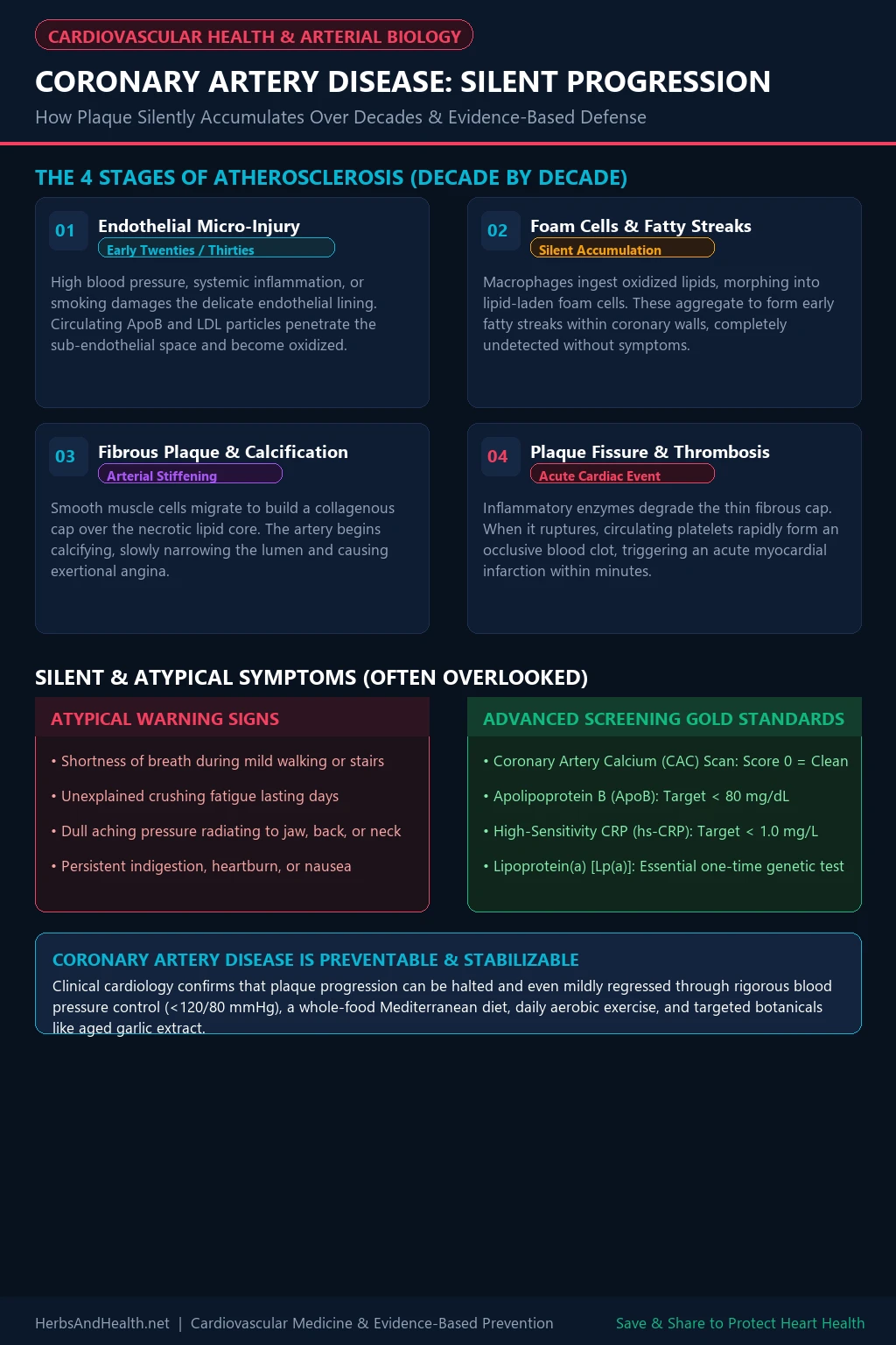
Save this visual progression guide to Pinterest or your phone to understand arterial plaque biology and share essential screening tools.
Silent and Atypical Warning Signs You Should Never Ignore
Hollywood movies have conditioned the public to believe that heart disease always announces itself through a theatrical “Hollywood heart attack”, someone clutching their chest in agonizing pain and falling to the floor. While crushing retrosternal chest pain radiating to the left arm certainly occurs, decades of epidemiological registry data reveal that nearly half of all heart attacks are “silent” or manifest with atypical, easily dismissed symptoms, particularly among women, older adults, and individuals with diabetes.
Be alert to these subtle red flags:
- Exertional Dyspnea (Shortness of Breath): Becoming unusually winded during activities you once handled with ease, such as walking up a flight of stairs or carrying groceries, is often an early indicator of myocardial ischemia. When coronary arteries are narrowed, the left ventricle stiffens under exertion, causing fluid pressure to back up into the lungs.
- Unexplained Chronic Fatigue: Experiencing overwhelming physical exhaustion that does not improve with sleep, especially when performing basic routine tasks, often reflects compromised cardiac output.
- Radiating Aches to Jaw, Neck, or Back: Cardiac pain fibers share neurological pathways with somatic sensory nerves in the spinal cord. Coronary ischemia frequently manifests as a dull, heavy ache in the jaw, lower neck, shoulder blades, or upper back rather than sharp chest pain.
- Persistent Dyspepsia and Nausea: Burning sensations in the upper abdomen, persistent acid reflux that does not respond to antacids, and unexplained nausea can signal ischemia along the inferior wall of the heart, which rests directly against the diaphragm.
For an in-depth breakdown of how acute cardiovascular crises differ, review our clinical comparison on heart attack versus cardiac arrest symptoms.
Beyond Standard Panels: Advanced Screening Gold Standards
Relying entirely on a standard fasting lipid panel and resting ECG provides a false sense of security. Nearly 50 percent of all heart attack victims present with “normal” calculated LDL cholesterol levels. When evaluating cardiovascular risk, modern preventative cardiology relies on four advanced objective diagnostic markers:
- Coronary Artery Calcium (CAC) Scan: A rapid, non-invasive, low-radiation CT scan that directly visualizes and quantifies calcified atherosclerotic plaque in your coronary arteries. A CAC score of zero indicates an extremely low 10-year risk of cardiovascular events, whereas scores above 100 or 400 indicate established, significant atherosclerosis requiring intensive preventive therapy.
- Apolipoprotein B (ApoB): Standard LDL tests measure cholesterol concentration within particles, but ApoB measures the exact particle count of all atherogenic lipoproteins (LDL, VLDL, IDL). Every atherogenic particle carries exactly one molecule of ApoB. An optimal primary prevention target is below 80 mg/dL (or below 60 mg/dL for high-risk patients).
- High-Sensitivity C-Reactive Protein (hs-CRP): A systemic inflammatory biomarker reflecting vascular endothelial stress. Plaque rupture rarely occurs in the absence of active vascular inflammation; target levels below 1.0 mg/L.
- Lipoprotein(a) [Lp(a)]: A genetically determined, highly atherogenic variant of LDL that also promotes thrombosis. Because Lp(a) levels are predominantly determined by genetics rather than diet, every adult should test Lp(a) at least once in their lifetime.
To explore how to naturally lower atherogenic particle concentrations, read our guide on natural remedies for cholesterol control.
Clinical Arterial & Cardiovascular Support Tools
Vetted nutritional tools supporting endothelial integrity, arterial compliance, and mitochondrial heart energy:
Kyolic Aged Garlic Extract Formula 100
Organic aged garlic extract rich in water-soluble S-allyl cysteine. Clinically proven in UCLA trials to slow coronary artery calcification, reduce soft plaque volume, and lower oxidized LDL.
Jarrow Formulas Ubiquinol QH-absorb 100mg
Active, bioavailable ubiquinol CoQ10 that fuels cardiac mitochondrial ATP synthesis, protects myocardial tissue from ischemic strain, and replenishes CoQ10 depleted by statin medications.
Doctor’s Best High Absorption Magnesium
Chelated magnesium lysinate glycinate that relaxes arterial smooth muscle cells, promotes endothelial nitric oxide synthesis, and maintains pliable blood vessels.
Evidence-Based Prevention: 5 Daily Habits to Protect Your Arteries
Atherosclerosis is neither inevitable nor irreversible. Intensive lifestyle intervention can halt plaque progression, stabilize vulnerable necrotic cores, and enhance myocardial perfusion. Implement these five foundational habits:
1. Adopt a Mediterranean Whole-Food Dietary Pattern
The landmark PREDIMED trial demonstrated that a Mediterranean diet rich in extra virgin olive oil and tree nuts reduces cardiovascular events by approximately 30 percent. Olive oil’s primary polyphenol, oleocanthal, functions as a natural COX inhibitor that reduces endothelial inflammation, while monounsaturated oleic acid resists oxidation within the vessel wall. Pair this with leafy greens, wild salmon, and legumes while minimizing ultra-processed seed oils and refined carbohydrates.
2. Maintain Strict Blood Pressure Control
Chronic hypertension mechanically batters the vascular endothelium, promoting micro-fissures where atherogenic particles lodge. Keep your resting blood pressure strictly below 120/80 mmHg. Prioritize high-potassium whole foods (spinach, avocado, bananas) to counterbalance sodium and promote vasodilation.
3. Engage in Regular Aerobic Exercise for Nitric Oxide Synthesis
Accumulate at least 150 minutes of moderate aerobic exercise (brisk walking, swimming, cycling) per week. Aerobic movement generates laminar shear stress against endothelial cells, triggering endothelial nitric oxide synthase (eNOS). Nitric oxide is your body’s master vasodilator: it keeps arterial walls supple, inhibits platelet aggregation, and prevents white blood cells from sticking to plaque sites.
4. Incorporate Targeted Cardioprotective Botanicals
Aged garlic extract has demonstrated remarkable efficacy in double-blind, randomized trials conducted at the Harbor-UCLA Medical Center, slowing the progression of coronary calcification and reducing low-attenuation plaque (the dangerous soft lipid core most prone to rupture). Pair aged garlic with standardized plant sterols to inhibit intestinal cholesterol absorption as detailed in our guide on how to lower LDL cholesterol naturally.
5. Eliminate Tobacco and Secondhand Smoke Exposure
Inhaled combustion chemicals trigger immediate endothelial dysfunction, oxidize circulating lipids, and dramatically thicken blood viscosity. Quitting smoking halts endothelial oxidative damage immediately; within one year of cessation, excess coronary risk drops by 50 percent.
Frequently Asked Questions About Coronary Artery Disease
Can existing arterial plaque actually be reversed?
Yes, to a meaningful degree. While densely calcified fibrous plaque rarely disappears completely, clinical trials utilizing intravascular ultrasound (IVUS) demonstrate that aggressive lipid lowering (achieving ApoB < 60 mg/dL), strict blood pressure control, and anti-inflammatory lifestyle habits can deplete the soft lipid-rich necrotic core, shrink plaque volume by 5 to 10 percent, and calcify the fibrous cap to prevent rupture.
What does a zero score on a Coronary Artery Calcium (CAC) scan mean?
A CAC score of zero means no calcified atherosclerotic plaque was detected in your coronary arteries. In asymptomatic patients, a score of zero confers an exceptional cardiovascular prognosis over the subsequent 5 to 10 years (often termed the “10-year warranty period”), with less than a 1 percent annual risk of a coronary event.
Why do some people with normal cholesterol have heart attacks?
Standard cholesterol panels measure total cholesterol content within particles, which can mask elevated particle counts (high ApoB). Additionally, cholesterol is merely one component of plaque; active vascular inflammation (hs-CRP), elevated Lipoprotein(a), and uncontrolled hypertension are powerful independent drivers of plaque formation and rupture even with normal cholesterol numbers.
What is the difference between stable angina and a heart attack?
Stable angina is predictable chest pressure that occurs during physical exertion or emotional stress and resolves completely with rest or sublingual nitroglycerin within five to ten minutes. A heart attack occurs when a plaque completely ruptures, forming a clot that blocks blood flow permanently; pain is severe, persistent, and accompanied by diaphoresis, shortness of breath, and nausea.
Is chest pain the only symptom of coronary artery disease?
No. In up to 40 percent of patients, especially women and individuals with diabetes, CAD presents as exertional shortness of breath, profound unexplained fatigue, or aching in the jaw, neck, or back without any classic chest pain. Any exertional discomfort between the jaw and navel warrants cardiovascular evaluation.
Peer-Reviewed Clinical References & Evidence
- Libby, P. (2002). Inflammation in atherosclerosis. Nature, 420(6917), 868-874.
- Budoff, M. J., et al. (2016). Aged garlic extract reduces coronary artery calcification: a randomized, double-blind study. Journal of Nutrition, 146(2), 433S-439S.
- Ference, B. A., et al. (2017). Low-density lipoproteins cause atherosclerotic cardiovascular disease: evidence from genetic, epidemiologic, and clinical studies. European Heart Journal, 38(32), 2459-2472.
- Estruch, R., et al. (2018). Primary prevention of cardiovascular disease with a Mediterranean diet supplemented with extra-virgin olive oil or nuts. New England Journal of Medicine, 378(25), e34.
- Hecht, H. S., et al. (2017). SCCT/STR guidelines for coronary artery calcium scoring of noncontrast noncardiac chest CT scans. Journal of Cardiovascular Computed Tomography, 11(1), 74-84.