Epidemiological research confirms up to 80% of adults experience lower back pain during their lifetimes, ranging from acute lumbar muscle spasms to persistent disc aches.
Because the spine comprises 33 vertebrae, discs, and nerves, back pain arises from diverse biomechanical drivers. Below is our clinical guide detailing how to distinguish muscle strains from disc herniations, identify urgent red flags, and implement the 5 core pillars of natural spinal rehabilitation.
📋 Medical Summary & Clinical Key Takeaways
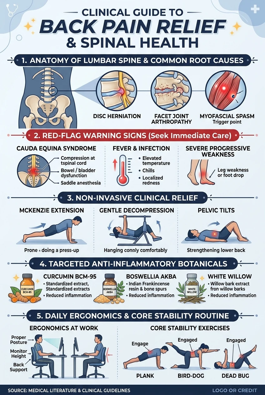
- Clinical Epidemiology: Up to 80% of adults experience lower back pain (lumbago), with over 90% of cases stemming from mechanical myofascial strain rather than permanent structural disc pathology.
- Red Flag Triage: Sudden loss of bowel or bladder control, saddle anesthesia, progressive lower-limb neurological weakness, or unexplained fever demand emergency evaluation for Cauda Equina Syndrome.
- Conservative Rehabilitation: Landmark spinal biomechanics research supports McGill’s “Big 3” core stabilization exercises (Curl-Up, Side Plank, Bird Dog) over aggressive spinal flexion or prolonged bed rest.
- Botanical & Nutritional Synergy: Bioavailable curcuminoids, boswellic acids, and elemental magnesium glycinate significantly reduce cyclooxygenase-2 (COX-2) inflammatory mediators and nocturnal muscle spasms.
Red Flags: When Back Pain is a Medical Emergency
While the vast majority of back pain episodes stem from benign muscular or mechanical issues that resolve with conservative care, certain spinal presentations indicate acute nerve compression, severe infection, or vascular emergencies. Seek immediate Emergency Room evaluation if your back pain is accompanied by any of the following red-flag symptoms:
- Loss of Bowel or Bladder Function (Cauda Equina Syndrome): Incontinence or urinary retention signals acute cauda equina compression requiring emergency surgical decompression within 24–48 hours to prevent permanent paralysis.
- “Saddle Anesthesia”: Numbness across the groin, buttocks, or inner thighs represents a surgical emergency.
- Progressive Motor Weakness: Sudden “foot drop” or severe lower-limb motor weakness indicates active spinal nerve root impingement.
- Unexplained Fever, Chills, or Night Sweats: May signal vertebral osteomyelitis, discitis, or a spinal epidural abscess.
- Cancer History with Intractable Pain: Unrelenting nocturnal bone pain unaffected by recumbency warrants urgent oncological evaluation.
Mechanical vs. Structural: Common Causes of Back Pain
Orthopedic specialists classify back pain into mechanical (involving muscles, ligaments, and joints) and structural/neuropathic (involving spinal discs and nerve roots):
1. Acute Lumbar Muscle & Ligament Strain
Accounting for over 70% of all acute back pain episodes, strains occur when the fibrous muscle bands (such as the erector spinae) or stabilizing ligaments supporting the lumbar curve are stretched beyond their mechanical threshold. Heavy lifting with rounded posture, sudden twisting, or coughing during awkward movements can trigger microscopic muscle tears, leading to intense localized swelling and protective muscle spasms.
2. Herniated or Bulging Intervertebral Discs
Intervertebral discs act as hydraulic shock absorbers between spinal bones. When the tough outer ring (annulus fibrosus) weakens, the gel-like interior (nucleus pulposus) can bulge or rupture outward. If this protruding material physically compresses or chemically irritates the adjacent spinal nerve root—most commonly at L4-L5 or L5-S1—it triggers sciatica: sharp, shooting, electric pain that radiates down the buttock, hamstring, and past the knee into the calf and foot.
3. Lumbar Spinal Stenosis
Most common in adults over age 50, spinal stenosis involves an age-related narrowing of the central spinal canal or neuroforaminal openings, typically driven by thickened ligaments and osteoarthritis bone spurs. People with stenosis characteristically experience leg heaviness and aching when walking or standing upright, but find rapid relief when bending forward or leaning on a shopping cart (the “shopping cart sign”), which temporarily widens the spinal canal.
4. Sacroiliac (SI) Joint Dysfunction
The sacroiliac joints connect your lower spine to your pelvis. When these dense joints become inflamed (sacroiliitis) or hypomobile due to uneven leg mechanics, pregnancy hormones, or repetitive impact, they generate sharp, stabbing pain localized to one side of the lower back, often centered over the posterior pelvic dimples.
5. Facet Joint Arthropathy
The facet joints are the small, cartilage-lined hinge joints connecting adjacent vertebrae. Chronic wear and tear erodes this cartilage, leading to bone-on-bone friction. Facet joint pain typically feels worse when leaning backward (lumbar extension) or twisting, and feels relieved when sitting or curling forward.
The 5 Core Pillars of Natural Back Pain Relief
Unless severe structural pathology or red-flag compression is present, conservative, non-invasive therapies yield recovery rates exceeding 90% within 4 to 8 weeks. Here are the 5 science-backed pillars of spinal rehabilitation:
Pillar 1: Active Recovery Over Strict Bed Rest
For decades, well-meaning doctors told back pain sufferers to lie flat in bed for weeks. Modern biomechanical science has completely dismantled this advice. Studies prove that bed rest beyond 48 hours actively delays recovery, causing rapid multifidus muscle atrophy, connective tissue stiffness, and increased subjective disability.
Instead, adopt active rest. Short, frequent, gentle walking sessions on flat terrain (10 to 15 minutes, 3 times daily) promote blood circulation through avascular disc tissues, flush out inflammatory metabolic byproducts, and prevent secondary muscular spasms.
Pillar 2: The McGill “Big 3” Stabilization Protocol
Developed by world-renowned spine biomechanist Dr. Stuart McGill, the “Big 3” exercises build muscular endurance in the torso without loading the vulnerable lumbar spine with dangerous flexion or compression. Clinical biomechanical trials published in Medicine & Science in Sports & Exercise (PubMed PMID: 12704689) confirm that these stabilization patterns maximize torso endurance while maintaining safe, sub-injurious spinal joint loads. Deep unilateral aching frequently stems from severe spasms in the deep stabilizing flank muscles; explore our clinical protocol on how to unlock your QL muscles for lasting back relief:
- The Modified Curl-Up: Lie on your back with hands under your lumbar arch. Lift head and upper shoulders 1 inch for 8 to 10 seconds, activating the rectus abdominis without compressive lumbar flexion.
- The Side Plank: Supported on elbow and knees or feet, elevate hips into a straight line. This activates the quadratus lumborum (QL) and lateral obliques without compressive shear.
- The Bird-Dog: Position yourself on hands and knees with a flat, neutral spine. Slowly extend your right arm forward and left leg backward until parallel to the floor, holding for 8 seconds without letting your pelvis tilt or sag. Switch sides. This activates the erector spinae and gluteus maximus without spinal shear force.
Pillar 3: The Strategic Heat vs. Cold Protocol
Knowing when to reach for an ice pack versus a heating pad makes a profound difference in symptom management:
- Cold Therapy (First 24 to 48 Hours): Apply an ice pack wrapped in a thin towel for 15 to 20 minutes every 2 hours following an acute injury. Cold constricts local blood vessels, blunting the acute inflammatory cascade and numbing hyperactive pain receptors.
- Moist Heat Therapy (After 48 Hours & Chronic Aches): Once acute swelling subsides, transition to moist heat. Heat dilates blood vessels, increasing oxygen delivery, soothing tense muscle knots, and restoring elasticity to tight connective tissues. Use heat for 20 minutes before gentle stretching or physical activity.
Pillar 4: Botanical Anti-Inflammatory Allies
Rather than relying indefinitely on synthetic NSAIDs (like ibuprofen), which carry risks of gastric ulceration and renal stress with long-term use, several standardized botanical extracts offer robust, targeted relief:
- Curcumin (Turmeric Extract): Curcumin inhibits inflammatory COX-2 and NF-kB pathways. Systematic clinical evaluations published in BMC Complementary and Alternative Medicine (PubMed PMID: 29315865) confirm that standardized curcuminoids paired with boswellic acids significantly reduce musculoskeletal pain scores and systemic inflammation comparable to standard non-steroidal anti-inflammatories without gastric ulceration risks. To further restore mobility and downregulate pain hypersensitivity, incorporate mindful stretching from our guide on yoga and natural healing remedies. Clinical trials show that high-absorption curcumin extracts (paired with piperine or phospholipid phytosomes) match the analgesic efficacy of low-dose pharmaceutical NSAIDs without gastrointestinal damage.
- Boswellia Serrata (Indian Frankincense): Rich in 3-O-acetyl-11-keto-beta-boswellic acid (AKBA), Boswellia selectively blocks the 5-LOX enzyme, curtailing inflammatory leukotrienes that drive cartilage erosion and joint stiffness.
- White Willow Bark (Salix alba): The ancient botanical precursor to modern aspirin, willow bark delivers natural salicin alongside protective flavonoids, offering long-lasting, gentle analgesia.
- Topical Capsaicin & Arnica Montana: Topically applied capsaicin depletes substance P (the neurotransmitter responsible for relaying pain signals to the brain), while homeopathic arnica reduces muscular bruising and localized tension.
Pillar 5: Targeted Micronutrients for Nerve & Bone Health
Your musculoskeletal system relies on precise nutritional cofactors to maintain neuromuscular tone and repair connective tissue:
- Magnesium Glycinate (300–400 mg daily): Competes with calcium in muscle fibers to ease nocturnal spasms and calm nervous system irritability with superior gastrointestinal tolerance.
- Vitamin D3 & Vitamin K2: D3 optimizes calcium absorption while K2 (MK-7) directs minerals into the vertebral bone matrix and away from soft tissue.
- Methylated B-Complex (B1, B6, B12): Supports nerve myelin sheath regeneration, dampening radiating tingling sensations associated with sciatic nerve irritation.
Sleep Posture & Workplace Ergonomics Checklist
You spend a third of your life in bed and another third in a chair. Optimizing these two daily postures is paramount for protecting your spinal curve. Because prolonged keyboard work frequently provokes concurrent upper-extremity repetitive strain, consult our ergonomic guide to wrist pain relief and carpal tunnel prevention:
| Setting | Optimal Setup | What to Avoid |
|---|---|---|
| Side Sleeping | Place a firm, contoured pillow between your knees to keep your hips and pelvis in neutral horizontal alignment. | Curling into a tight fetal ball or allowing the top knee to collapse onto the mattress. |
| Back Sleeping | Place a medium bolster or folded pillow beneath your knees to relax tension on the psoas and lumbar spine. | Using oversized neck pillows that thrust your cervical spine forward. |
| Stomach Sleeping | Avoid entirely. If essential, place a thin flat pillow beneath your pelvis to reduce lumbar sway. | Flat stomach sleeping twists the cervical spine 90 degrees and hyper-extends lumbar facets. |
| Desk Ergonomics | Adjust your chair so your knees and hips rest at 90-degree angles with feet flat on the floor; monitor at eye level. | Slumping forward, perching on the edge of the seat, or crossing legs for extended periods. |
| The 30-Minute Rule | Stand up, stretch, and perform 5 gentle standing lumbar extensions every 30 to 45 minutes of seated work. | Remaining seated continuously for hours without posture changes. |
Recommended Back Pain Relief Essentials
These three third-party tested, ergonomic and nutritional tools provide clinically supported, home-based recovery support:
1. Doctor’s Best High Absorption Magnesium
100% chelated with TRAACS magnesium glycinate and lysinate. Superior bioavailability to relax tight, spasming spinal muscles, reduce nocturnal cramping, and promote restorative, restorative sleep without digestive upset.
2. Penetrex Joint & Muscle Therapy Cream
Fast-acting, non-greasy topical relief cream infused with arnica, vitamin B6, MSM, and boswellia serrata. Delivers soothing, targeted relief directly to aching lower back muscles, tendons, and facet joints without burning scents.
3. Everlasting Comfort Memory Foam Lumbar Pillow
Ergonomically contoured 100% pure memory foam cushion with dual adjustable straps. Fits office chairs and car seats to reinforce natural lumbar lordosis and prevent slouching-induced disc compression.
Related Clinical & Musculoskeletal Guides
- Unlock Your QL Muscles: Relieve Deep Lower Back Tension Naturally
- Yoga & Mind-Body Healing: Science-Backed Movement & Herbal Synergy
- Wrist Pain Relief: Root Causes, Carpal Tunnel Tests & Ergonomics
- What is Eczema? Causes, Common Types, Triggers & Natural Barrier Relief
- Dry, Itchy Skin Relief: Causes, Lipid Barrier Science & Natural Remedies
Frequently Asked Questions (FAQ)
Can applying a heating pad make back inflammation worse?
Yes, if applied during the initial 24 to 48 hours when local blood vessels are dilated and inflammatory mediators are surging. Heat during this early window can aggravate swelling. Use cold therapy for the first 48 hours, transitioning to soothing moist heat once acute inflammation stabilizes.
How can I tell if my back pain is coming from my kidneys or my spine?
Kidney pain originates higher in the back—under the lower ribs on either side of the spine (the flank area). It typically presents as a steady, deep, sickening ache that does not change whether you bend, twist, or lie down, and is often accompanied by fever, painful urination, or blood in the urine. Spinal and muscular pain, by contrast, sharpens with movement, twisting, or lifting, and eases with supportive resting positions.
Is walking really good for a herniated disc?
Yes. Gentle, upright walking decompresses the lumbar spine, promotes circulation, and drives fluid imbibition through intervertebral discs without high-impact rotational forces.
When should I insist on getting an MRI for my back?
According to American College of Physicians guidelines, routine MRI imaging is not recommended for uncomplicated back pain within the first 6 weeks, as scans frequently show incidental bulges that do not correlate with pain. An immediate MRI is indicated if red-flag symptoms (Cauda Equina, progressive neurological weakness, cancer history) are present, or if severe pain persists without improvement past 6 weeks of structured conservative care.
How long does a typical back muscle strain take to heal?
Most uncomplicated lumbar muscle strains show marked improvement within 2 to 4 weeks when managed with active recovery, gentle mobility, and proper ergonomics. Full tissue remodeling and ligamentous stability typically solidify by 6 to 8 weeks.
Medical Disclaimer: This guide is intended for informational and educational purposes only and does not replace personalized medical evaluation, diagnosis, or treatment. If you experience severe, worsening, or radiating back pain, consult a board-certified spine specialist, orthopedic physician, or licensed physical therapist.